Principles for the diagnosis of prostate adenoma (benign prostatic hyperplasia)
This entry was made in order to help figure out why doctors prescribe certain examinations for adenoma.
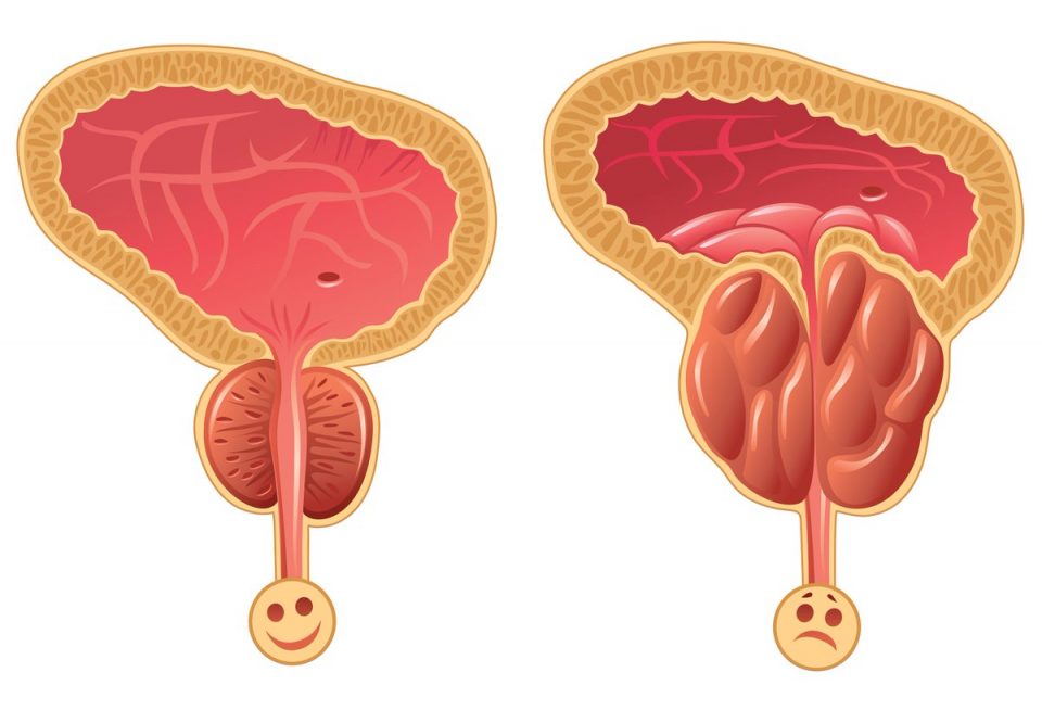
Why is prostate adenoma dangerous? With this disease, the urethra is compressed, the process of urination is disrupted, and the outflow of urine worsens. Therefore, it is very important for the doctor to conduct an initial diagnosis and determine the size, location and effect of the adenoma on urination.
For this, first of all, three surveys are carried out, which I will tell you about in more detail.
The first is a transrectal ultrasound study, according to the results of which it becomes clear whether, in principle, there is adenomatous tissue and what size it is. There are cases when a large prostate gland does not cause any clinical manifestations and is accidentally detected during routine examinations. In other cases, the medium-sized prostate gland causes severe clinical manifestations: difficulty urinating, a sluggish stream of urine, a feeling of incomplete emptying.
When conducting an ultrasound scan, the doctor sees the location of the adenomatous (altered) tissue relative to the urethra. According to this parameter, two variants of the development of events can be distinguished: it is located along the entire urethra and in this case, even with small sizes, causes a pronounced compression of the canal. Or the growth is more in the peripheral part of the prostate gland, the urethra is only partially affected, and even with a significant size, the symptoms may be less pronounced or even absent.
The next stage of diagnosis is the assessment of urethral compression. For this, a study such as uroflowmetry is performed. The study is carried out with a moderately full bladder. Urination is carried out in a special device, while the speed indicators of the stream are assessed. The conclusion is issued in the form of graphs, which for certain diseases and conditions have their own characteristics, which help the doctor to assess the work of the urinary system.
The bladder https://en.wikipedia.org/wiki/Urinary_bladder is a hollow muscular organ that is supposed to relax (store urine) and contract (evacuate urine). If the work of the bladder during contraction is difficult due to the compression of the urethra, the bladder begins to work in an enhanced mode, involving its compensatory mechanisms for this.
The clinical manifestation of adenoma of the prostate gland just appears at the moment when the bladder cannot adequately contract, since all compensatory mechanisms have been spent.
Conducting an ultrasound of the bladder in this situation, we see a change in the walls of the bladder, as well as the presence of residual urine, which indicates the impossibility of adequate emptying (contraction) of the bladder. This is another necessary examination for patients with this problem.
Focusing on the results of the examinations, the doctor selects the tactics of management or one or another treatment regimen.